Mastering the Method: A Systematic Approach to Veterinary Abdominal Ultrasound
Jul 13, 2023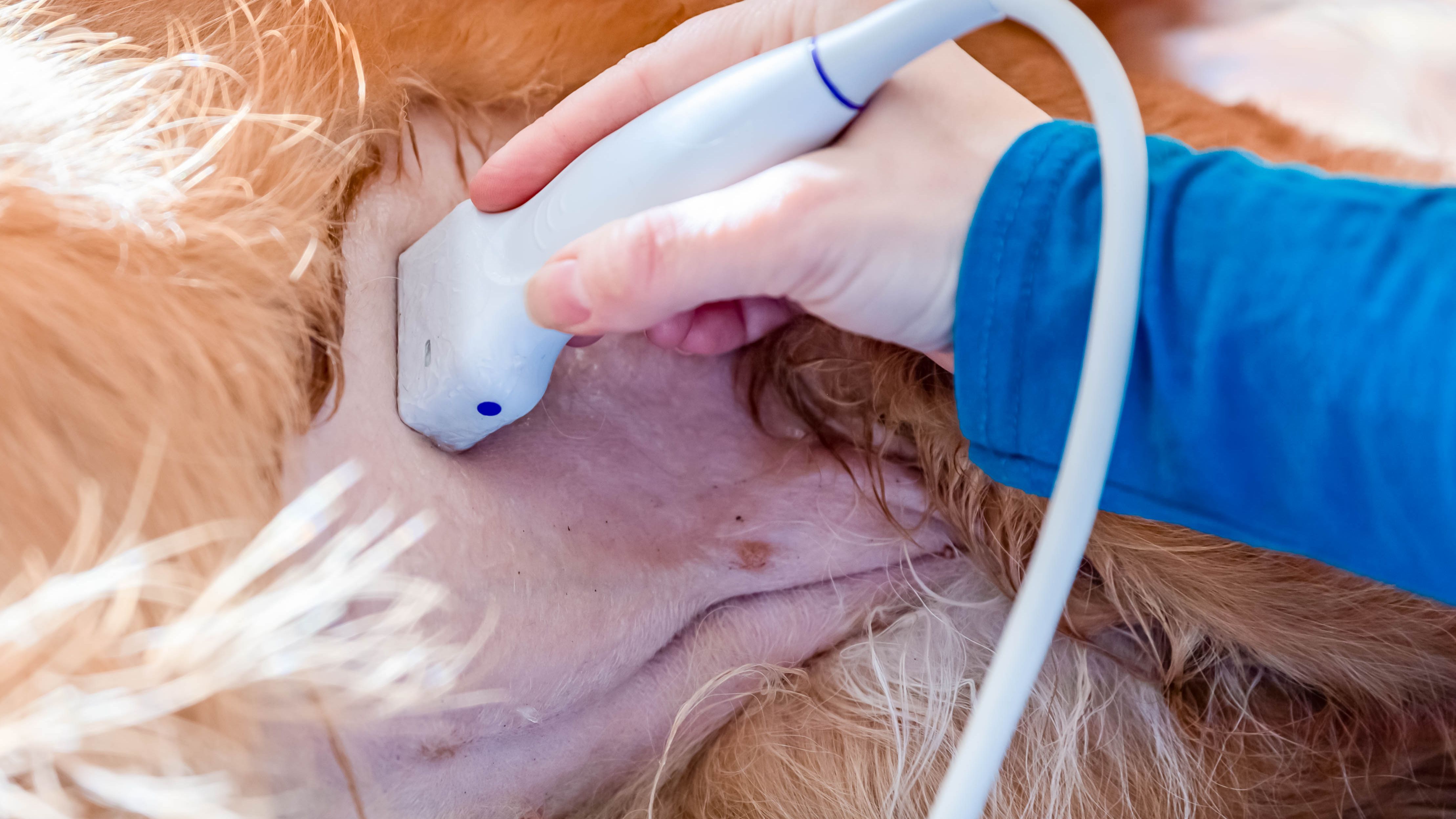
Introduction
Something I frequently hear from veterinarians is their struggle to recognise what "normal" looks like during an ultrasound examination. In this blog post, I'm here to provide some advice and share a systematic approach that will help you gain a clearer picture of normal and enhance your ability to spot pathology.
Consistency is Key
When it comes to recognising what's normal, consistency is key. By scanning more frequently and establishing a consistent routine, you'll become increasingly familiar with the baseline appearances of various abdominal structures. This familiarity is essential for identifying any abnormalities that may arise.
The FOVU Method
A Step-by-Step Guide: To facilitate a systematic and comprehensive approach to abdominal ultrasound, I recommend following this method. Let's break it down:
- Right Lateral Recumbency: Begin with the animal in right lateral recumbency, focusing on the left-hand side of the abdomen.
- Liver, Stomach, Spleen, and Left Kidney: Start just behind the xiphisternum, examining the liver. Move up the costal arch to visualize the stomach, spleen, and left kidney.
- Aorta, Left Medial Iliac Lymph Node, and Left Adrenal Gland: Follow the aorta, observing the left medial iliac lymph node and then proceeding back to the left adrenal gland.
- Jejunum, Bladder, Colon, and Left Limb of the Pancreas: Continuing the scan, assess the jejunum, bladder, colon, and then move cranially to examine the left limb of the pancreas.
- Left Lateral Recumbency: Flip the animal over to left lateral recumbency to scan the right-hand side of the abdomen.
- Gallbladder and Urinary Bladder: Begin by examining the gallbladder, followed by the urinary bladder, checking for any movement or sediment.
- Right Kidney, Right Medial Iliac Lymph Node and Right Adrenal Gland: Trace the caudal vena cava to the right medial iliac lymph node and then to the right adrenal gland.
- Duodenum, Right Limb of the Pancreas, Jejunal Lymph Nodes, Ileum, and Ileocaecocolic Junction: Next, evaluate the duodenum and pyloroduodenal junction. Proceed to the right limb of the pancreas, jejunal lymph nodes, ileum, and ileocaecocolic junction.
Maintaining The Flow
The beauty of this method is its flow. By learning and consistently applying this approach, you can seamlessly move from one organ to another without removing the probe from the animal. This efficient method allows for quicker recognition of abnormalities and facilitates a more accurate assessment of the entire abdomen.
Conclusion
By adopting a systematic approach like the method I’ve described during your abdominal ultrasound examinations, you'll rapidly gain confidence in identifying normal structures and swiftly detect any deviations from the norm. So, embrace the method, repeat it with each scan, and watch as your ability to recognise normal from abnormal becomes second nature.
Master the method, master the ultrasound exam!
